Anatomical Specification: Cubital Fossa
Region: Anterior aspect of the elbow joint.
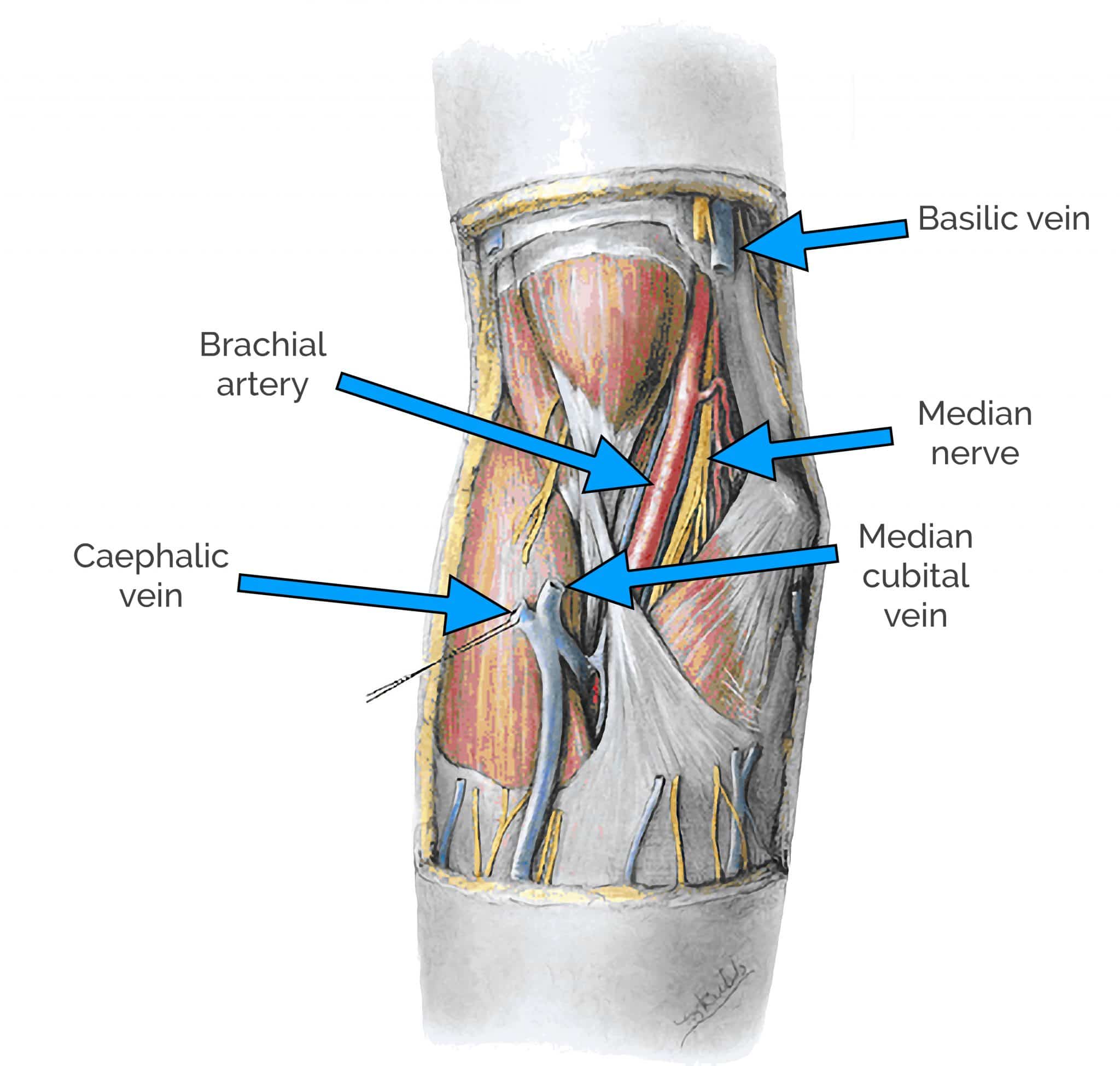
Description: The cubital fossa is a triangular anatomical space or depression located on the anterior surface of the elbow. It serves as a significant passageway for major neurovascular structures as they transition from the arm to the forearm.
Boundaries:
Superior (Proximal): An imaginary line connecting the medial and lateral epicondyles of the humerus.
Lateral: Medial border of the brachioradialis muscle.
Medial: Lateral border of the pronator teres muscle.
Floor: Brachialis and supinator muscles.
Roof: Skin, superficial fascia (containing the median cubital vein, medial and lateral cutaneous nerves of the forearm), and bicipital aponeurosis.
Contents (from medial to lateral, typically):
Median Nerve: Innervates most of the anterior compartment muscles of the forearm.
Brachial Artery: Divides into the radial and ulnar arteries just distal to the fossa.
Biceps Tendon: Inserts into the radial tuberosity.
Radial Nerve (deep branch): Divides into superficial and deep branches within or just distal to the fossa.
Clinical Significance:
Venipuncture: The median cubital vein, located in the roof of the fossa, is a common site for blood draws and intravenous injections due to its accessibility and superficial position.
Blood Pressure Measurement: The brachial artery in the cubital fossa is where the stethoscope is placed to listen for Korotkoff sounds during blood pressure measurement.
Nerve Entrapment: Conditions like pronator teres syndrome can involve compression of the median nerve within this region.
Pulse Palpation: The brachial pulse can be palpated in the cubital fossa.
Function: Facilitates the passage and protection of vital nerves and blood vessels crucial for the function of the forearm and hand.